Patients have been concerned about the radiation risks from CT scans for a long time. This is because a CT scan can expose a person to 70 to 200 times more radiation than a regular X-ray.
However, there’s some good news. CEA-Leti, a research institute sponsored by the French government, has created photon modules. These modules, when used with a new type of CT scanner being developed by Siemens Healthineers (a division of Siemens AG), have shown to decrease the amount of radiation a person would be exposed to. This is a significant step towards making CT scans safer.

CT scans use a kind of radiation known as ionizing radiation, which is the same type of radiation we’re exposed to naturally every day. Experts say that we’re exposed to about 3 millisieverts (mSv) of this radiation each year. A sievert is a unit that measures the effect of low levels of ionizing radiation on the human body.
A CT scan can expose a person to between 1 and 10 mSv of radiation, depending on the radiation dose and the part of the body being scanned. For example, a low-dose chest CT scan exposes a person to about 1.5mSv, while a regular dose exposes them to about 7mSv.
CEA-Leti, a research institute, has developed a photon module for CT scans that can reduce the amount of X-ray exposure to patients. This module, known as an X-ray photon-counting detector module (PCDM), has been shown in clinical trials to improve image quality by reducing image noise and distinguishing multiple contrast agents, and it could potentially increase spatial resolution.
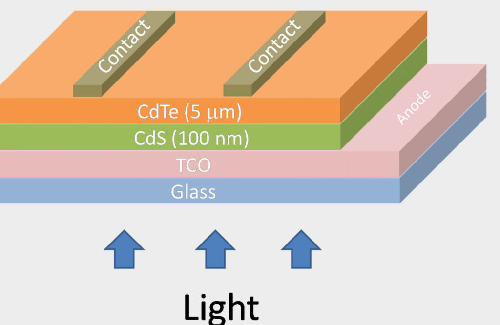
These PCDMs are made from a stable crystalline compound called cadmium telluride (CdTe), which is often used in solar cells and as an infrared optical window. It’s usually combined with cadmium sulfide to create a type of solar cell called a p–n junction solar photovoltaic cell.

Typically, CdTe PV cells use a structure known as n-i-p, which allows for the capture of high-resolution and multi-energy images at the same time. High-resolution images are made possible by using a detector with small pixels. The multi-energy feature provides color images, unlike the grey-scale images produced by traditional detectors, and allows for the accurate identification of the atomic number of any chemical elements in the body.
X-ray CT scanners work by using a computer to process many X-ray measurements taken from different angles to create cross-sectional images of the object being scanned. Current X-ray CT scanners use energy-integrating detectors (EIDs), which are based on a technology that indirectly converts X-ray photons into electronic signals. First, the X-ray photons are converted into visible light using a material called a scintillator. Then, the visible photons produce electronic signals using a device called a photodiode. In contrast, photon-counting detector modules (PCDMs) convert X-ray photons directly into electronic signals, which is a more efficient process.
EIDs record the total energy deposited in a pixel over a certain period of time, without distinguishing between low and high-energy photons. This results in a black-and-white X-ray image that shows the density of the body’s organs. PCDMs, on the other hand, count each photon, which improves the contrast-to-noise ratio of the image. The energy classification of the detected photons can be used to produce a color image that allows for the accurate identification of the atomic number of any chemical elements and can identify multiple contrast agents present in the body.
Finally, the detector module’s very high spatial resolution produces clearer images of extremely fine structures, such as small airways in the lungs, trabeculae in bones, and thin wires in coronary stents, than current scanner technology.
“The idea of Siemens Healthineers to integrate PCDMs in the future generation of X-ray CT scanners was new, and no available technology existed when CEA-Leti began working on this,” said Loick Verger, the industrial-partnership manager for X-ray imaging at CEA-Leti.
Verger explained that CEA-Leti used its simulation tools to design the geometry of the detector, chose a semiconductor-based on CdTe, designed the electronic readout circuit, and then was able to propose a reliable CdTe electronic assembly technology.
“The idea of Siemens Healthineers to integrate PCDMs in the future generation of X-ray CT scanners was new, and no available technology existed when CEA-Leti began working on this,” said Loick Verger, the industrial-partnership manager for X-ray imaging at CEA-Leti.
Loick Verger from CEA-Leti explained that they used simulation tools to design the shape of the detector, selected a semiconductor based on CdTe, designed the electronic readout circuit, and then proposed a reliable assembly technology using CdTe.
In a separate study, researchers at the Mayo Clinic in the United States tested the performance of Siemens Healthineers’ photon-counting detector system in phantoms, cadavers, animals, and humans. The images produced from over 300 patients consistently showed the theoretical benefits of this type of detector technology, leading to several important clinical advantages.
Cynthia McCullough, a professor of Medical Physics and Biomedical Engineering at the Mayo Clinic, explained that their research papers have shown improved image sharpness, reduced radiation or iodine contrast dose requirements, and decreased levels of image noise and artifacts.
It’s clear that any reduction in the amount of X-ray radiation we’re exposed to is beneficial and reduces the risk of changes in our body’s cells. It’s natural for us to be concerned about radiation exposure, and making CT scans safer is definitely a positive development. However, it’s important to keep things in perspective. Studies in the US looking at the health risks of CT scan radiation have concluded that you’re 24 times more likely to die in a road accident than from an illness caused by CT scan radiation.

